×

; ?>)
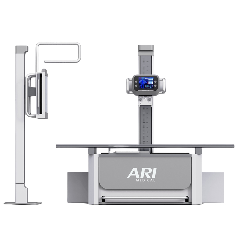
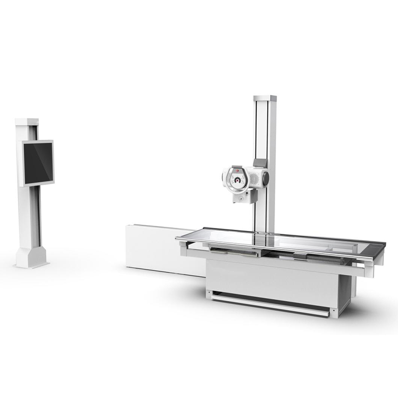
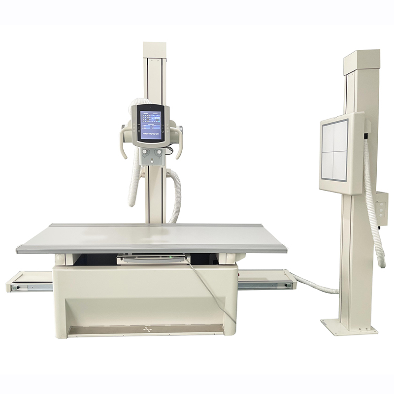
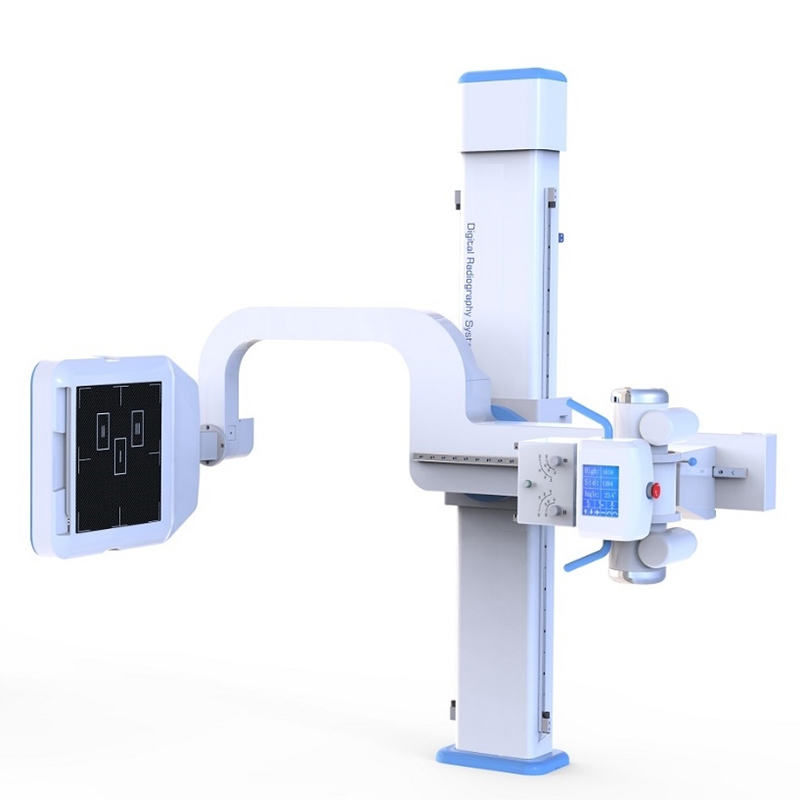
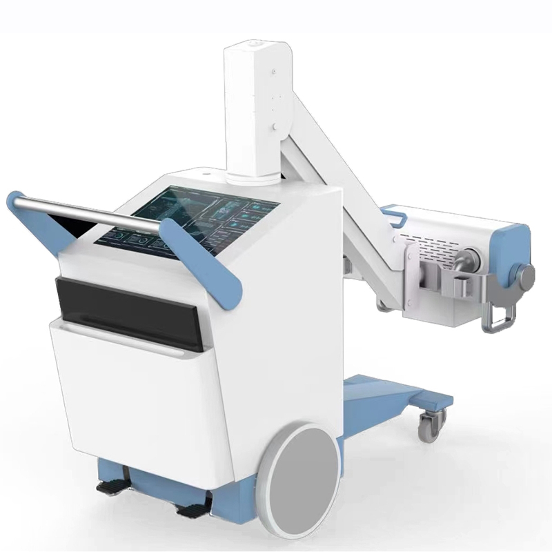
The machine is Combined X-ray photography of medical diagnostic equipment, application for wards, emergency rooms, operating rooms, ICU, etc. It can be used for human body head, limbs, chest, waist and abdomen and many other parts. It provides finest image resolution through Flat Panel detector. Easy operations make the work efficient
| PARAMETERS | SPECIFICATION |
| X-ray Generator | |
| Output power | 32KW |
| Frequency | ≧30KHZ |
| Current | 10~400mA |
| Voltage | 40~125KV |
| mAs | 0.1~320As |
| Exposure time | 1ms~10000ms |
| Intelligent protection system | It has self-protection system. It will give an alarm and check fault code. |
| X-ray Tube | |
| Anode type | Rotating Anode |
| Focal Spot value | 1.0mm /2.0mm |
| Power of focal spot | 21kW/42.5kW |
| Maximum voltage | 125KV |
| Revolving speed of anode | 2800rpm |
| Inherent filtration | 1.5mmAl |
| Anode heat storage capacity | 100kJ(140kHU) |
| Heat storage capacity | 900kJ |
| Anode angle | 16° |
| Maximum anode heat dissipation | 475W |
| Flat Panel Detector | |
| Type | Amorphous Silicon WIFI connection |
| Scintillator | Cesium Iodide |
| Active Area | 14x17Inch(35cm*43cm) |
| Active Pixel | 2304*2800 |
| Pixel Pitch | ≦150μm |
| A/D Conversion | 16bits |
| DQE | ≧70% |
| Spatial Resolution | 36Lp/cm |
| Acquisition time | ≤2S |
| AED function | Equipped |
| Collimator | |
| Type | Manual Collimator |
| Max. Window | 440mm×440mm(SID=100cm) |
| Lamp | AC/DC24V、5W |
| Lamp timer | Automatic illumination with timer for lamp(30S) |
| Inherent filtration | 1.0mmAl |
| Illuminant | LED |
| The tube bracket structure | |
| X-ray tube support way | Folding arm |
| Tube focus distance to the ground | 480mm~1700mm,±5% |
| Arm’s Folding Range | -45~+45° |
| Tube Tilting Angle | ±45° |
| Tube Vertical Rotation Angle | 360° |
| Collimator axial rotation | ±360° |
| Digital image workstation | |
| Display size | 19 inch LCD touch screen |
| Display screen Maximum
resolution |
1280*1024 |
| CPU | ≥2.0GHz |
| Memory | ≥2GB |
| HDD | ≥320GB |
| OP System | 64 bit Windows operation system |
| DICOM3.0 | Query for integration with any PACS |
| Functions | Patient registration: acquisition, find the patient data, to prepare for the photography |
| Radiographic examination:setting system parameters,
exposure photography and get the image |
|
| Images display, image analysis, image processing and output | |
| Data management: managing patient data, leading in, leading out , batch file, etc in accordance with DICOM3.0 | |
| Configure calibration: the system components such as flat-panel detector,such as tube for automatic configuration and calibration | |