×

; ?>)
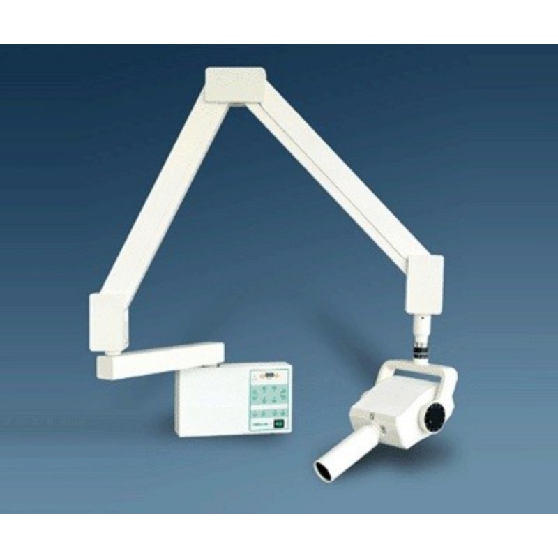
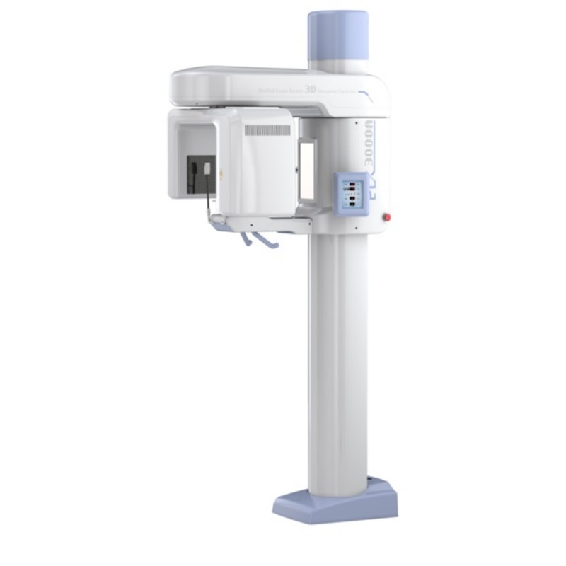
Oral and maxillofacial cone beam computed tomography equipment (dental CT) with 12cmX15cm flat panel detector, can be widely used in oral and maxillofacial surgery, orthodontics, orthognathic surgery, implant, dental, temporomandibular joint Preoperative and postoperative.
1, Using the world’s top high-end flat panel detector, high resolution, small distortion, uniform brightness.
2, Use exposure mode, only when needed to activate the X source. During the rotation of the 18s, the rays last only 4-8s. The effect of reducing the check dose, reached the international advanced level.
3, 3000A proprietary three-dimensional reconstruction algorithm, the original two-dimensional sequence projection into a three-dimensional volume image, you can at any angle, any location to provide high-definition tomography.
4, Through the three-dimensional volume image extraction of oral panorama, can provide high-definition panoramic images.
5, The image can be used to generate reports, print or save as a report file.
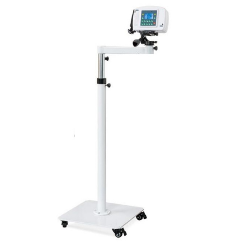
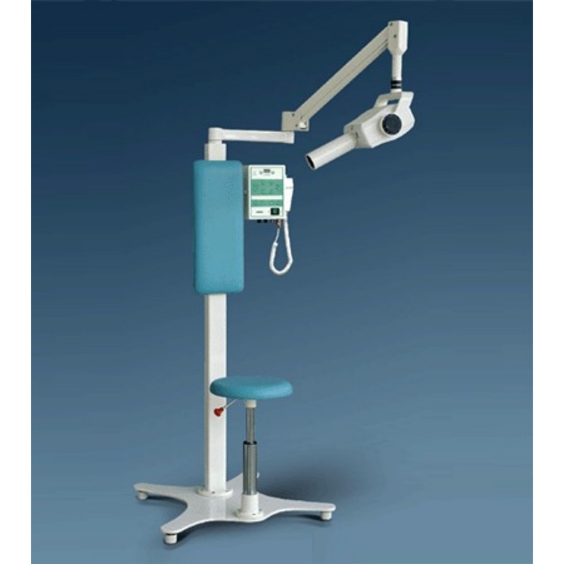
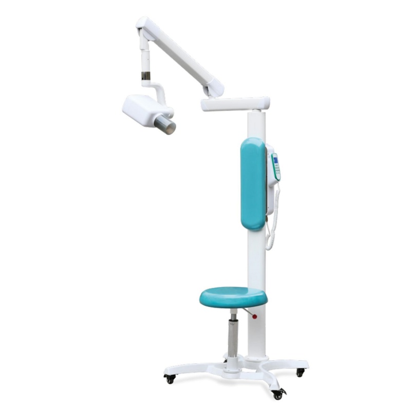
6, Can provide standing, sitting and wheelchair and other kinds of film.