×

; ?>)
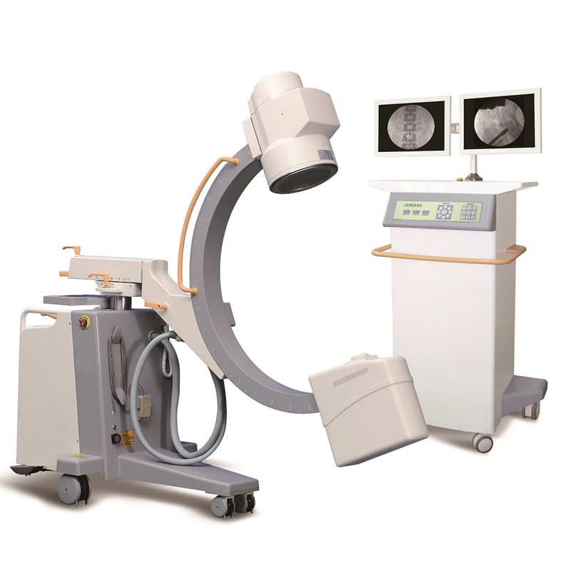
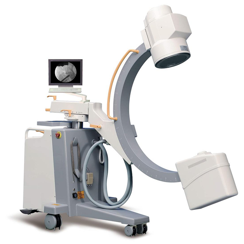
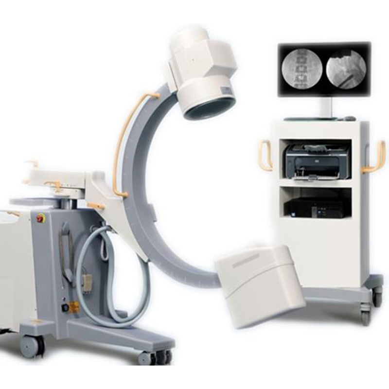
Widely use in operating room, X-ray department, emergency room, intervention and peripheral angiography.
General surgery, urology, chest surgery, trauma, spine surgery, abdominal surgery, orthopedics, bone reduction and fixation, endoscopy and surgery. Intervertebral disc angiography and angioplasty.
-With HF & HV generator, inverter, and High quality image intensifier, the x ray generator can work stably. It also reduces the damage for the doctors caused by radiation.
-10.4″ LCD friendly interface control board: easy operation and can be rotated to 180° (L90°, R90°). Compared with ordinary key board, our LCD display is not easy to be oxidized.
-Pulse and boost fluoroscope provides all kinds of size of patients with clear diagnosis.
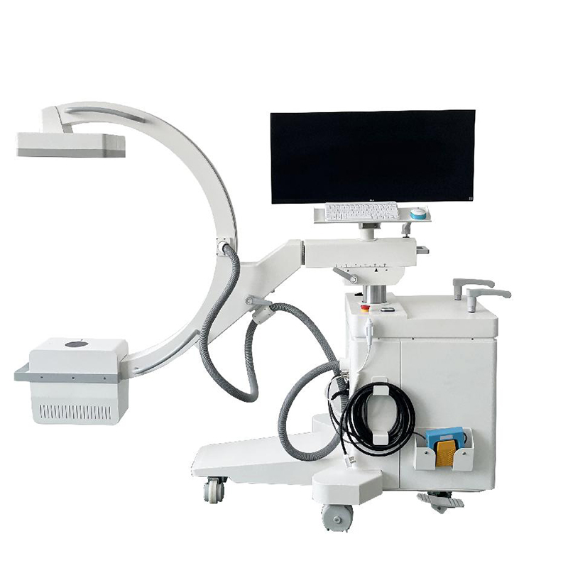
-With a steering handle, it can adjust the direction quickly when moving. And parallel movement function is also available.
-Equipped with unique block line wheels, it can avoid accident when the machine moves.
-Large free space in arc: the space can reach 800mm, which is convenient for operation.
-9″/6″/4.5″ three fields of vision: it facilitates diagnosis of partial enlargement.
-CCD camera with rotation function: range of camera rotation: 360°.
-Original Korean linear slide rail can make the the movement of the machine flexibly.
-Applied with Omron Relay, the noise can be reduced and the life time of the equipment can be increased.
| PARAMETER | SPECIFICATION |
| HF generator | 120KV, 5KW, 40kHz |
| Fluoroscopy mode | Fluoroscopy mode: 40~120kV, 0.5~5mA (KV manual and automatic);
Pulsed fluoroscopy: 40~120kV, 0.5-5mA Boost fluoroscopy: 40~120kV, 5~10mA; Photography: 40~120kV, 1~250mAs |
| X ray tube | Dual-focus Rotating anode: 0.3mm/0.6mm
Italy IMD Technology |
| Image intensifier | Three fields of vision 9, 6, 4.5, Japan Toshiba Brand |
| CCD camera | Japanese Watt Brand, 360° rotation |
| Image system | 5 Degree recursive Noise Reduction
Frame freezing Thousands of image data storage Image conversing and rotating Screen display with image comparison Support “one click” print, print the image report Equip with Dicom 3.0 network port (optional) Support Dicom 3.0 standard data export function |
| Mechanical motion specifications | Vertical travel: 0-400mm
Horizontal travel: 0~200mm Rotation about horizontal axis: ±180° |
| C-arm dimension | Distance of focal spot to image generator(SID): 1000mm
Depth in arm: ≥650mm Mobile stand: 1800mm*800mm*1850mm Image system: 750mm*530mm*1680mm |
| Standard configuration | High resolution CCD camera: 1set
x-ray generator: 1set 9” image intensifier: 1set image processor 24” LCD high definition display: 1 set Mobile stand: 1 set Printer: 1set |
| Dimension | Two cases: A) 252×93×174cm
B) 90×80×140cm G.W.: 500KG |
| NO. | DESCRIPTION |
| 1 | With HF & HV generator,inverter,and High quality image intensifier, the x ray
generator can work stably. It also reduces the damage for the doctors caused by radiation. |
| 2 | 10.4″LCD friendly interface control board: easy operation and can be rotated to 180°
(L90°,R90°) Compared with ordinary key board, our LCD display is not easy to be oxidized Besides, the trouble will be displayed in English, which will improve the efficiency of fault diagnosis. |
| 3 | With a steering handle, it can adjust the direction quickly when moving.And parallel
movement function is also available. |
| 4 | Equipped with unique block line wheels, it can avoid accident when the machine moves. |
| 5 | Thales brand Image intensifier: 9″/6″/4.5″ three fields of vision:it facilitates diagnosis of
partial enlargement. |
| 6 | Original Korean linear slide rail can make the movement of the machine flexibly. |
| 7 | Applied with Omron Relay, the noise can be reduced and the life time of the equipment can be increased. |
| 8 | Japanese Watt Brand CCD camera with rotation function: range of camera rotation:360°. |
| 9 | Pulse and boost fluoroscope provides all kinds of size of patients with clear diagnosis. |
| 10 | Large free space in arc: the space can reach 800mm,which is convenient for operation. |