×

; ?>)
| Project | Model | Features and Performance |
| High Voltage Generator
(MOS Technology) |
DTXR-50 | Input: 110v/220v/380v,
Output power: 50kw, Inverter frequency >300KHz, Tube Current:10~500mA, Current time product: 1mAs~630mA, Tube Voltage: 40~125KV, Can be directly connected to the network cable remote control for remote fault diagnosis and maintenance. |
| X-ray tube
(CANON) |
E7242X | Dual-focus 0.6mm/1.5mm, power 20/46KW,
maximum current 500mA, maximum voltage125KV, Heat storage capacity 200KHU. |
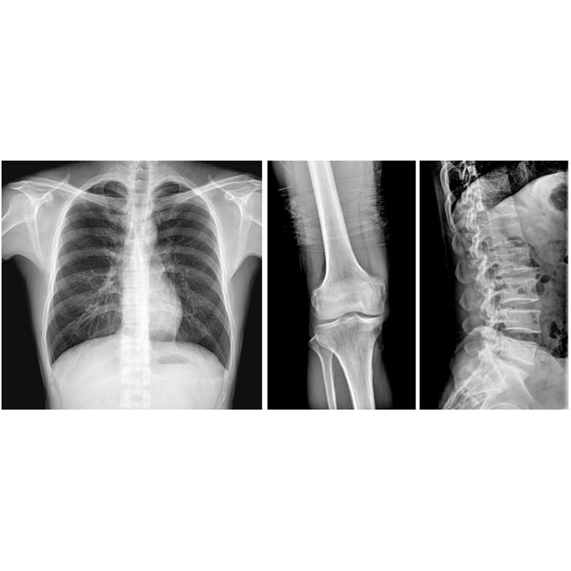
| Flat Panel Detector | 1800L | Strontium iodide amorphous silicon panel.
Pixel matrix > 3072 x 3072; Imaging area: 430x430mm” Pixel size: 140μm, Spatial resolution: 3.5Lp/mm, AD conversion 16bits; MTF:> 60% @1lp/mm> 20% @3lp/mmDQE:(@RQA5, 30μGy)>65% @0lp/mm >20% @3lp/mm Dynamic Range: >80dB Imaging time: The output time of the original image is less than 3 seconds, and the post-loading software processing should be about 8 seconds. |
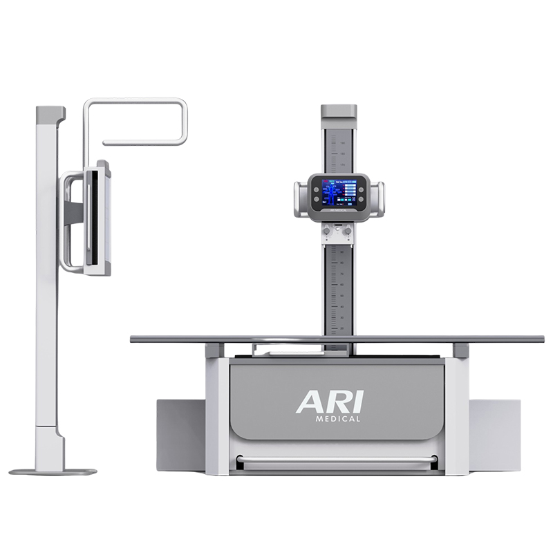
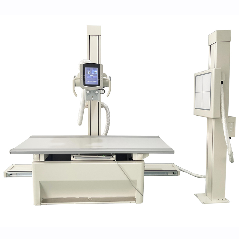
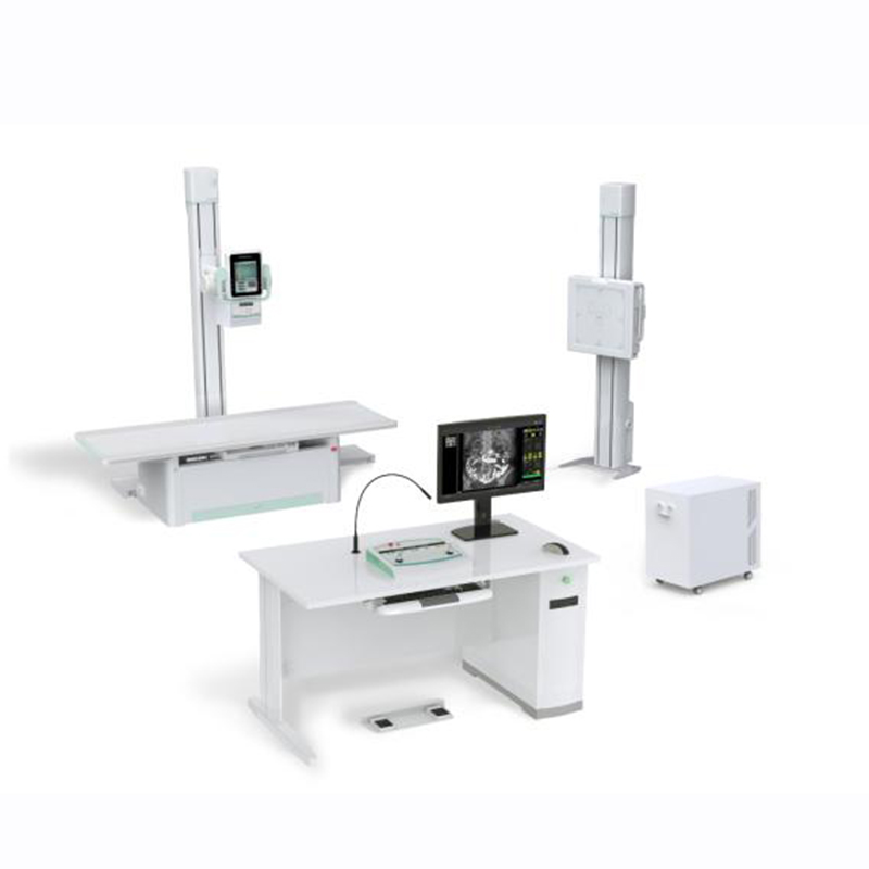
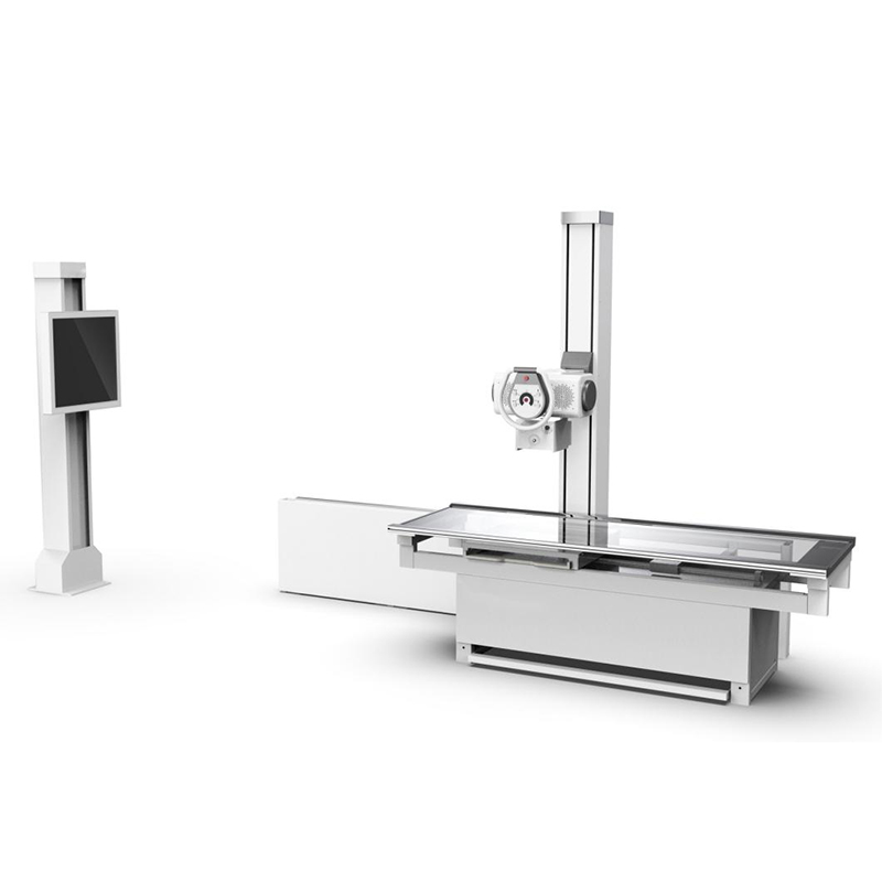
| Photograph Bed | SYC-2 | Detector longitudinal motion range: 1000mm,
Table lateral movement 180 mm. Bed panel material: polycarbonate polymer panel, Mechanical Motion Control: Manual electromagnetic lock. SID1000mm, Korean vibrating grid. |
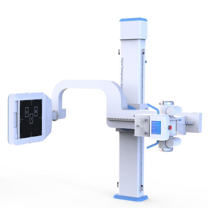
| Tube stand | ZJ-1 | The X-ray tube assembly moves up and down no less than 900mm;
Longitudinal movement is not less than 1800mm; X-ray tube rotates along the horizontal axis ≥ ± 180 ° |
| Collimator | XSQ-1 | The total number of deviations between the sides of the ray radiation field and the respective sides of the light field does not exceed 3% of the distance from the field plane to the focal point. The sum of the deviations of the two axes should not exceed 4% of the indicated focal point to the detector plane. |
| Photograph Stand | SYJ-1 | Detector activity: Vertical movement no less than 900mm;
SID1800mm, Korean vibrating grid |
| DR Image Processing
Software Workstation |
DR | BGD Sequential Interconnection Acquisition Software,
DICOM3.0 standard, pre-selection of projection site; preset image brightness/contrast, window width/window position; organization equalization technology preset; image enhancement and sharpening technology preset; image segmentation, transmission, printing, etc. Can be connected with PACS system. |
| Computer Host
DELL |
2011 | CPU 2GHz or more,
memory > 4G hard disk > 2000G, can store pictures in large capacity |
| Professional Diagnostic Display | ML2002 | Size: 21.3″; Type: Monochrome; Mpixel:2MP; Resolution: 1600×1200;
Max Brightness: 1900cd/m2; Grayscale tone:16bit. |
| Remote Box | Can be remote controlled tube stand and photography frame, suitable for large-size chest X-ray examination | |
| Operation display | Size: 19″; Brand:Dell |