
×

; ?>)
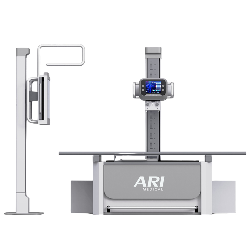
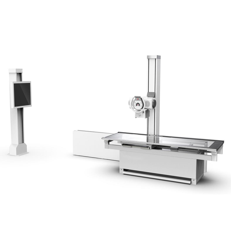
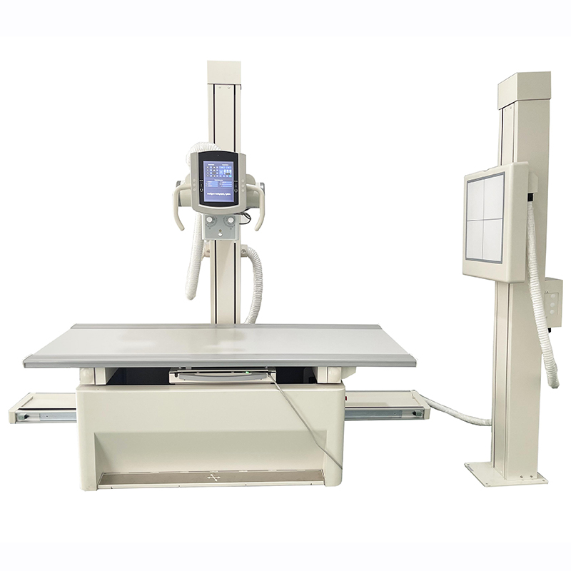
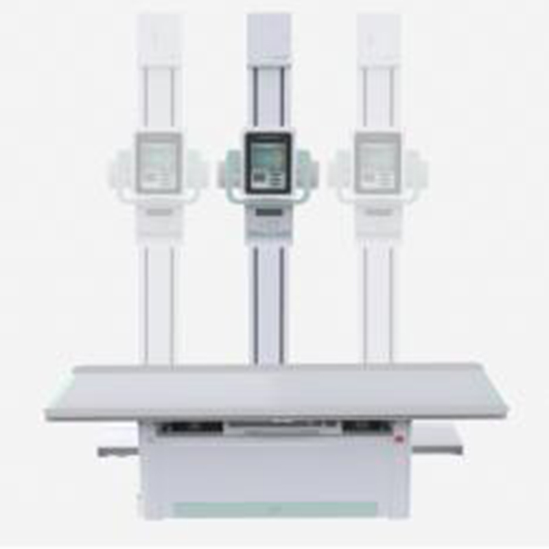
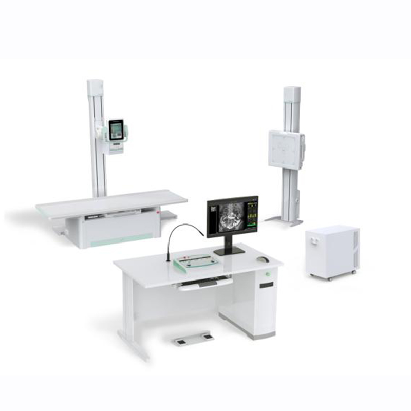
AR-7900I is a dual functional machine integrated digital radiography and digital fluoroscopy into one machine. It can work in radiology department where in Medical Center, Diagnostic Center, Health Examination Center, Orthopedics and Trauma for doing radiography and fluoroscopy exams like, Chest, limbs abdomen, lumbar etc whole body part radiography and fluoroscopy like esophagography, pyelography, salpingography, chest fluoroscopy, abdomen fluoroscopy etc.
Digital radiography:
Digital fluoroscopy:
Digital angiography :
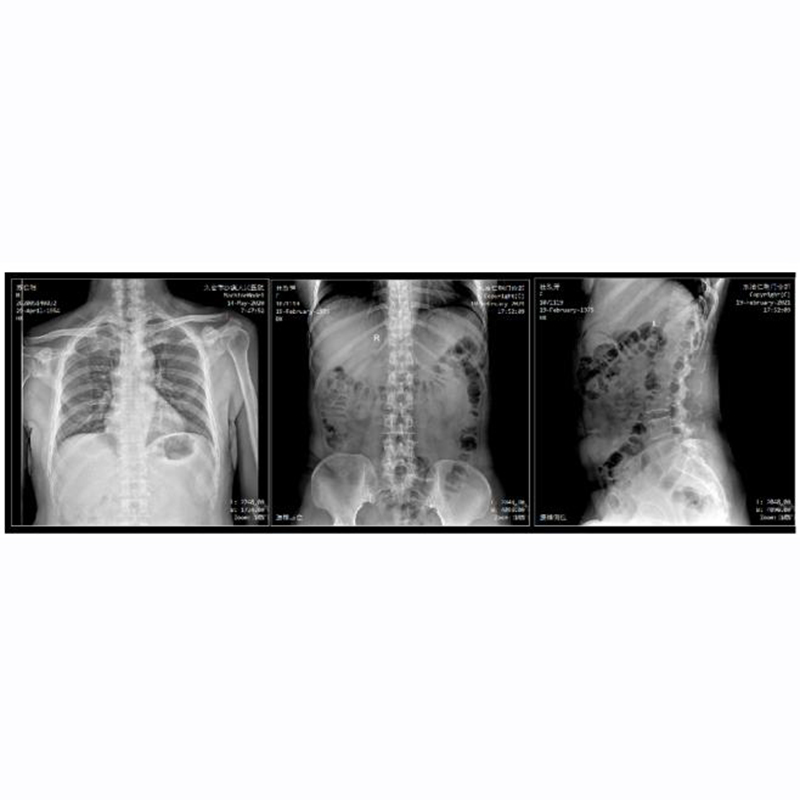
Precise Radiography:
Multi-dimensional Radiography:
Special Radiography:
Spine stitching Radiography:
| PARAMETER | CONTENT | SPECIFICATION |
| Power condition | Voltage | 380V±38V |
| Frequency | 50Hz±1Hz | |
| Capacity | ≥85kVA | |
| Internal Resistance | ≤0.17Ω | |
| HF HV Generator System | Power | 65.5KW |
| Inverter Frequency | 500 KHz ±20% | |
| Tube voltage | 40kV—150kV | |
| Tube current | 10mA—800mA | |
| mAs | 0.1mAs—800mAs | |
| Fluoroscopy tube voltage | 40kV—125kV | |
| Fluoroscopy tube current | 0.5mA~20mA (continuous) | |
| 5mA~40mA (pulse) | ||
| Touch screen Table-side control | 10.4 inch | Image display, SID stretching range, exposure parameters, x-beam size, head rotation angle, body type, and emergency examination |
| X ray tube (CANON E7252X) | Tube Focus:big/small | 1.2mm /0.6mm |
| Input power | Big focus: 75kW/Small focus 27kW | |
| Anode thermal capacity | 210kJ/300KHU | |
| X-ray tube heat capacity | 900kJ/1250KHU | |
| Rotary anode speed | 9700rpm | |
|
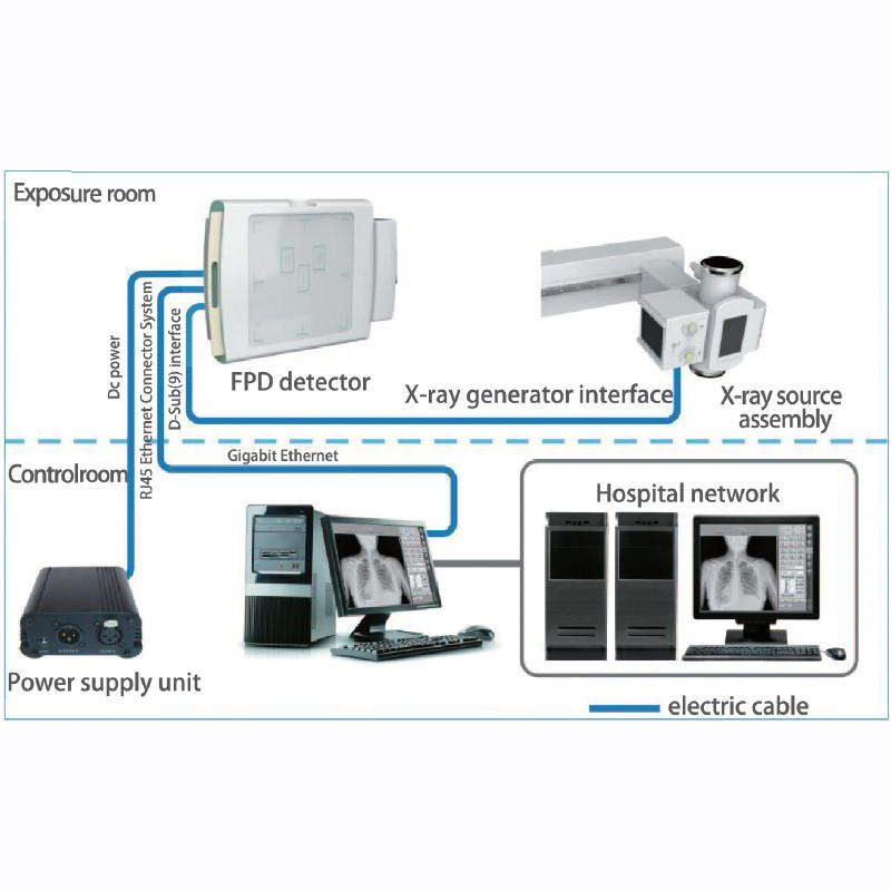
Digital Flat Penal Detector |
Active area | 427mm(H)×427mm(V) |
| Pixel matrix | 3072(H)×3072(V) | |
| Pixel pitch | 139 μm | |
| Limiting resolution | Min. 3.7 line pair/mm | |
| A / D transition | 16 bit | |
| Energy range | 40 – 150 kVp | |
| Acquisition speed | 30fp/s | |
| X-ray table | Light touch switch, electromagnetic brake | |
| Height | 550mm | |
| Load capacity | 200kg | |
| Filter (Grid) | Focusing distance: 100cm,
Grid density: 230L/INCH, |
|
| Movement | electromagnetic brake 4-way floating | |
| Mechanical movement | X-ray tube up-down | 1300mm |
| X-ray tube rotating | -90°~90° | |
| Pillar longitudinal movement | 1500mm | |
| Pillar rotating | -180°~180° | |
| Table longitudinal moving | 1000mm | |
| Table Transverse moving | 260mm | |
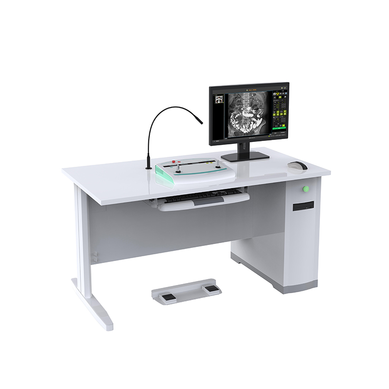
| Digital Workstation | 19inch monitor +computer +keyboard +mouse +speaker | |
| Joystick remote controller | ||
| Foot brake control system | ||
| Desk | ||
| Image professing software | ||
| Image professing software functions introduction :
1. Basic operations and image acquisitions
|
Registration: regular registration, emergency registration, adding agreement, adding items, clearing information, starting inspection
Work list: list information, search for patients to be examined, refresh of the to-be-examined list, deletion of examinations, and display column settings. Start inspection, emergency registration Exam list: list information, examined patient display and search, delete images, image storage, CD burning, add items, display column settings, modify examination information Patient size: thin adults, adults, fat adults Photography parameter settings: exposure mode, frame rate setting, kVp, mA, ms, mAs , AEC, focus selection Perspective parameter setting: exposure mode, frame rate setting, kVp, mA, ABS, time reset Browsing Tools: Zoom, Flip Horizontal, Flip Vertical, Rotate Left 90 Degrees, Rotate Right 90 Degrees, Zoom In, Zoom Out, Original Size, Move Image, Invert Color, Adaptive Size, ROI Magnifier, Magnifier, Default Window Width Window Level, ROI window width and window level, visible window width and window level, point gray value, advanced processing, ellipse gray measurement Measurement tools: arrow, cardiothoracic ratio (CTR), distance measurement, angle measurement, spine measurement System Tools: Text Marker, Anterior Body Marker, Left Marker, Right Marker, Circular Crop, Delete Image, Delete Tool, Error reset, Exposure Indicator, Full Screen, Save Current Image, Print |
|
| Image professing software functions introduction :
2. Image output and management operations:
|
Report editing: Patient information display and editing, image selection, report content template selection, report description, report conclusion, report description + conclusion, editing knowledge base, reporting doctor, reviewing doctor, reporting time, printing template, setting, saving report
Report Printing: Quick Print, Print Report Image Archive, Burn, Print: Delete Image, Image Storage, Browse Image, Report, Lock/Unlock, Store Queue, Print Queu Disc Burn: Volume Label, Save Settings, File Compression, File Structure Print: Access to DICOM Printer, Local Printer System Settings: System, Annotation Information, Tools, Others Hardware Configuration: Syncbox, High Voltage, Probe, Collimator, DAP Network Configuration: Local, Job List, Netstore, Local Store, Print Check Management: Basic information, placement information, hardware parameters, image processing, inspection protocol Quality management: Search, system management User management: Add, update, delete, authority |
|
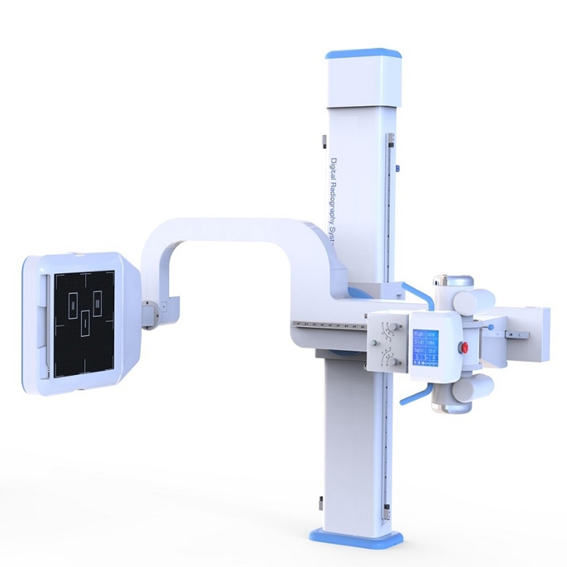
Flexible and efficient multi-dimensional movement
Full satisfaction in varieties of positions.
Cost-Effective:
Reliable & High Efficiency:
Clinically-Oriented:
Customer-Oriented:
Value-Added: