×

; ?>)
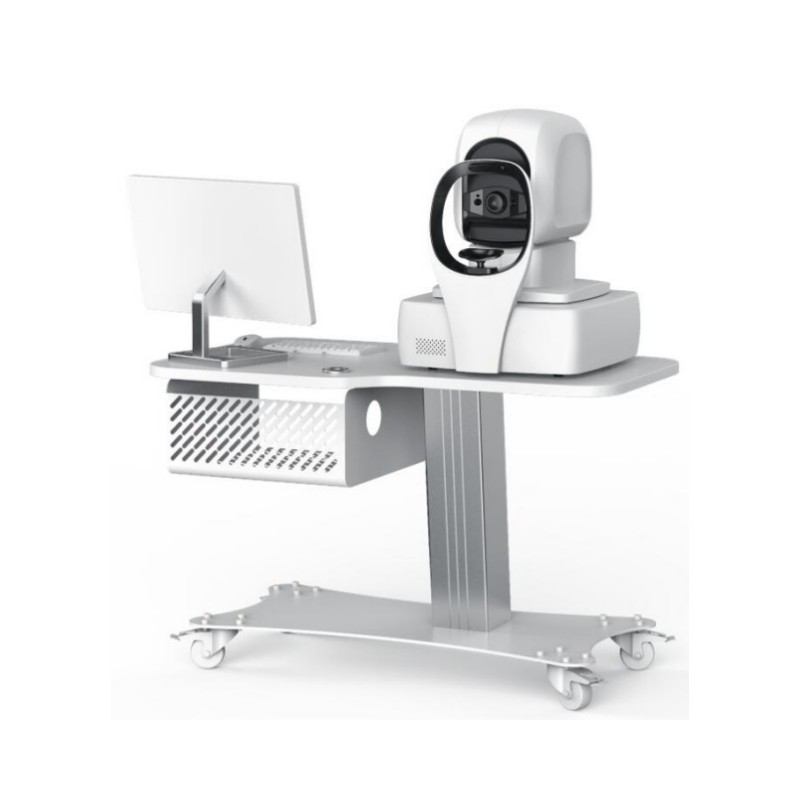
The OCT-1000 Optical Coherence Tomography for Ophthalmology is an integrated automatic artificial intelligence OCT system with both soft and hard capabilities, boasting complete independent intellectual property rights. It combines cutting-edge multi-image registration enhancement technology with industry-leading analysis techniques to offer a comprehensive solution for clinical diagnosis and treatment. This includes automated image capture, precise image analysis, intelligent and real-time cloud-based data sharing.
Ultra-fast fundus signal acquisition
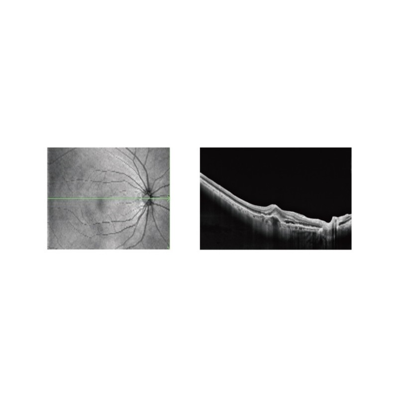
80,000 times per second “second” acceleration technology, the scanning speed is greatly improved, helping to accurately capture different subdivision forms of fundus lesions in a shorter time and obtain high-quality images.
Diagram of the lesion
It can be used for auxiliary diagnosis of 16 main fundus diseases
| ITEM | OCT-1000T | OCT-1000S | OCT-1000M |
| Type | SD-OCT | SD-OCT | SD-OCT |
| Light energy (cornea) | ≤750μW | ≤750μW | ≤750μW |
| Measurement characteristic | Axial resolution:5μm
Lateral resolution:20μm |
Axial resolution:5μm Lateral resolution:20μm |
Axial resolution:5μm Lateral resolution:20μm |
| Scanning characteristic | Maximum A-scan speed:≥20000times/second Scanning depth:2.3mm Maximum scanning range:12mm*9mm |
Maximum A-scan speed:≥80000times/second Scanning depth:2.3mm Maximum scanning range:12mm*9mm |
Maximum A-scan speed:≥55000times/second Scanning depth:2.3mm Maximum scanning range:12mm*9mm |
| Light Source characteristics | Central wavelength:840nm Optical power:≤750μW Refractive compensation range:-20D~+25D |
Central wavelength:840nm Optical power:≤750μW Refractive compensation range:-20D~+25D |
Central wavelength:840nm Optical power:≤750μW Refractive compensation range:-20D~+25D |
| Scanning mode | 5 kinds Large field of view: HD straight line, radiation six lines, local area: macula, optic disc |
7 kinds Large field of view: HD straight line, radiation six lines, area Local area: macula, optic disc Front section: HD straight line, radiation six lines |
7 kinds Large field of view: HD straight line, radiation six lines, area Local area: macula, optic disc Front section: HD straight line, radiation six lines |
| Scanning range | 12mm*9mm Complete the image acquisition of macula + optic disc at one time |
12mm*9mm Complete the image acquisition of macula + optic disc at one time |
12mm*9mm Complete the image acquisition of macula + optic disc at one time |
| Single line scan length | 12mm One scan macula+ optic disc |
12mm (Anterior segment 6mm) |
12mm (Anterior segment 6mm) |
| Pupil requirement | 2mm | 2mm | 2mm |
| Automatic pupil orientation | √ | √ | √ |
| Automatic pupil compensation | √ | √ | √ |
| Fundus image | Full pixel imaging technology based on high scanning speed | Full pixel imaging technology based on high scanning speed | Full pixel imaging technology based on high scanning speed |
| Focusing mode | Fully automatic | Fully automatic | Fully automatic |
| Software analysis |
Large field analysis Local analysis – Optic disc 8 items Local analysis – macular 8 items Anterior segment analysis (peripherals required) |
Large field analysis Simultaneously covering macula and optic disc a total of 14 itemsLocal analysis – Optic disc 8 items Optic disc 6*6mm fundus image Cup-disc ratio Six lines Horizontal C/D Ratio Vertical C/D Ratio Optic cup area RNFL ring sweep thickness curve RNFL thickness topographic map TSNI thickness grid of nerve fiber layer Local analysis – macular 8 items Anterior segment analysis (peripherals required) |
Large field analysis Simultaneously covering macula and optic disc a total of 14 itemsLocal analysis – Optic disc 8 items Optic disc 6*6mm fundus image Cup-disc ratio Six lines Horizontal C/D Ratio Vertical C/D Ratio Optic cup area RNFL ring sweep thickness curve RNFL thickness topographic map TSNI thickness grid of nerve fiber layer Local analysis – macular 8 items Anterior segment analysis (peripherals required) |
| User profile | √ | √ | √ |
| Integrated artificial intelligence | It can assist in the
diagnosis of 16 major ophthalmic diseases accurately identify, segment and automatically label the focal area |
It can assist in the
diagnosis of 16 major ophthalmic diseases, accurately identify, segment and automatically label the focal area |
It can assist in the
diagnosis of 16 major ophthalmic diseases, accurately identify, segment and automatically label the focal area |
| DICOM interface function | √ | √ | √ |
| Mode of operation | Fully automatic, manual operation | Fully automatic, manual operation | Fully automatic, manual operation |
| MODE | SCANNING WAYS | PHYSICAL SIZE | SLICE DIRECTION |
| Large field | Single-line | 12mm | Horizontal |
| Six radial lines | 9mm | Per 30° | |
| Large field | 12mm x9mm | horizontal or vertical | |
| Glaucoma | Macular area | 6mm x6mm | Horizontal |
| optic disc area | 6mm x 6mm | Horizontal | |
| Anterior segment | Single line | 6mm | Horizontal |
| Six radial lines | 6mm | Per 30° |