Search
Search
Enquiry List
0
Your enquiry list is empty, please kindly add products.
Send Enquiry
Home
About Us
Profile
Philosophy
Overview
Quality Commitment
Products
Solutions
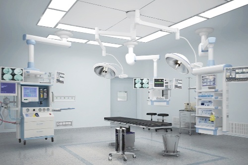
Operating Theatre
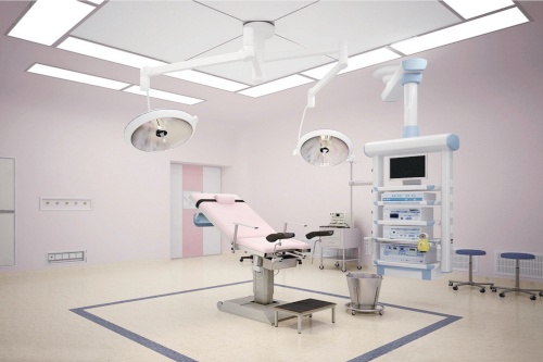
Delivery Room
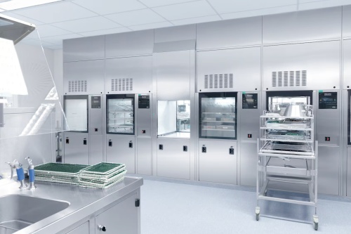
Sterilization Room
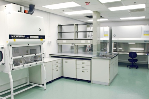
Laboratory
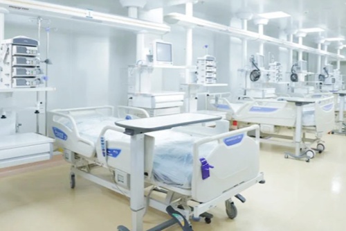
Ward
Dental Clinic
ENT Clinic
Veterinary Clinic
Services
2-Year Warranty
Service Policy
FAQ
Download
Cooperation
Seeking Distributors
OEM & ODM
Technical Cooperation
Suppliers Cooperation
News
Exhibition & Event
News Release
Contact
X
Search
Search
Close this search box.
Home
About Us
Profile
Philosophy
Overview
Quality Commitment
Products
Solutions
Operating Theatre
Delivery Room
Sterilization Room
Laboratory
Ward
Dental Clinic
ENT Clinic
Veterinary Clinic
Services
2-Year Warranty
Service Policy
FAQ
Download
Cooperation
Seeking Distributors
OEM & ODM
Technical Cooperation
Suppliers Cooperation
News
Exhibition & Event
News Release
Contact
Enquiry List
0
Previous slide
Next slide
Solutions
More
Operating Theatre
Delivery Room
Ward
Sterilization Room
Laboratory
Featured Products
More
AR-D3 Anesthesia Machine
ARQ DR X-ray System
EOT-2000 Operating Table (Electro-hydraulic)
AML700/700 LED Operating Lamp
DEF-7000 Defibrillator
AR-K10 4D Ultrasound Scanner
Products
More
Products
More
Operating Theatre Equipment
Anesthesia Machine
Ventilator
Defibrillator
Electrosurgical Unit
Patient Monitor
Medical Pendant
LED Operating Lamp
Operating Lamp
Operating Table
Delivery Table
Suction Unit
Syringe Pump
Infusion Pump
Feeding Pump
Blood Infusion Warmer
Operating Microscope
Shockwave Lithotripter
Surgical Tools
OB/GYN Equipment
Fetal Doppler
Fetal Monitor
Colposcope
Gynaecological Bed
Infant Incubator
Infant Radiant Warmer
Infant Phototherapy unit
Jaundice Meter
Infant Scales
Mammography X-ray System
Diagnostic Equipment
Vascular Doppler Detector
Ultrasound Scanner
Ophthalmic Ultrasound
ECG Machine
Holter ECG & Stress ECG
Bone Densitometer
EEG & EMG
Pulse Oximeter
Spirometer
Audiometer
Examination Lamp
Hand-Held ETCO2
Blood Glucose Monitor
Vein Finder
Sterilizer (Autoclave)
Washer Disinfector
Ethylene Oxide Gas
Portable Type
Table Type
Vertical Type
Horizontal Cylindrical Type
Pulse Vacuum Table Type
Pulse Vacuum Horizontal Type
H2O2 Low-Temp Plasma
Dry heat (Hot air) Type
Cassette Type
UV Sterilizer
Industrial Washing Machine
Laboratory Equipment
Biological Microscope
Spectrophotometer
High Speed Centrifuge
Low Speed Centrifuge
High Speed Freeze Centrifuge
Low Speed Freeze Centrifuge
Special Centrifuge
Thermostatic Bath
Laboratory Shaker
Laboratory Mixer
Water Distiller
Magnetic and Electric Stirrer
Laboratory Incubator
Dry Oven
PH Meter
Biological Safety Cabinet
Clean Workbench
Water Purifier
Ultrasonic Cleaner
Pipette
Microtome
Medical Hand Sink
Medical Freezer
Blood Bank Refrigerator
Medical Pharmacy Refrigerator
Analysis Balance
Medical Destroyer
X-ray Series
CT
DR X-ray System
C-arm X-ray System
Mobile X-ray Machine
Radiography X-ray System
Mammography X-ray System
Dental X-ray Machine
Portable X-ray Machine
X-ray Film Illuminator
X-ray Film Processor
X-ray Protective Products
In-Vitro Diagnostics
Specific Protein Analyzer
Immuno Fluorescence
Feces Analyzer
Biochemistry Analyzer
Coagulation Analyzer
Hematology Analyzer
Microplate Reader/Washer
Electrolyte Analyzer
Glycated Hemoglobin Analyzer
Blood Gas Analyzer
Urine Analyzer
PCR Analyzer
Hemodialysis Equipment
Hemodialysis Machine
Dialyzer Reprocessing Machine
R.O Water Purification Machine
Dialysis Consumables
Dialysis Chair
Hemodiafiltration Machine
Hospital Furniture
Manual Hospital Bed
Electric Hospital Bed
ICU Bed
Child Bed
Medical Trolley
Chairs
Bedside Cabinet
Patient Stretcher
Appliance Cupboard
Examination Couch
Overbed Table
Ward Equipment
Other
Bed Mattress
Walking Aids
Crutches and Canes
Walkers
Rollators
Bath Bench
Commode Seat
Commode Wheelchair
Steel Wheelchair
Aluminum Wheelchair
First-Aid Products
Defibrillator
Portable ventilator
Spine Board
Ambulance Stretcher
Scoop Stretcher
Folding Stretcher
Vacuum Stretcher
Basket Stretcher
Resuscitator
First aid bag
Stretcher Base
Stair Stretcher
Medical Human Model
Emergency Skills Training
Nursing Skills Training
Clinical Skills Training
Maternity Skills Training
Diagnosis Skills Training
Anatomy Model
Ophthalmic Equipment
Slit lamp
Ophthalmoscope&Retinoscope
Ophthalmic Table&Chair
Refractometer
Auto Lensmeter
Phorptor
Lens Edger
Auto chart Projector
Optometry Unit
PD Meter
Vision Chart
OCT
Biometer
Dry Eye Detector
Tonometer
Vision Screener
Perimeter
Fundus Camera
Phaco Emulsifier
Keratometer
Ophthalmic Ultrasound
Dental Equipment
Dental Unit
Dental Autoclave
Dental X-ray Machine
Dental Accessories
ENT Equipment
ENT Diagnostic Set
Otoscope
Fiber Laryngoscope
Video Laryngoscope
ENT Treating Desk
ENT Treating Chair
Home Care Equipment
Sphygmomanometer
Blood Pressure Monitor
Oxygen Concentrator
Nebulizer
Scales
Sleep therapy system
Bedsore Prevention Mattress
Veterinary Equipment
Veterinary Anesthesia machine
Veterinary ECG Machine
Veterinary Monitor
Veterinary Operating Table
Vet Ultrasound scanner
Vet Boold Pressure Monitor
Vet X-Ray Machine
Veterinary Pulse Oximeter
Veterinary Medical Pump
Medical Consumables
Surgical Staplers
Surgical Sutures
ESU Consumables
Medical Mask
Disposable Syringe
Medical Cannula
Medical Gloves
Sterilization Pouch
Chest Drainage System
Medical Endoscope
Gastroscope
Colonoscope
Ureteroscope
ENT Endoscope
Cystoscope
Choledochoscope
Bronchoscope
Duodenoscope
Hysterscope
Endoscope Washer
Personal Protective Equipment
Disposable Coverall
Forehead Infrared Thermometer
Face Shield
Eye Protector
Rapid Test Kit
Face Mask